Orthopediquize

🍁 Widespread orthopedic diseases,diagnosis and treatments
🍁 Surgical procedure videos

Community chat: https://t.me/hamster_kombat_chat_2
Website: https://hamster.network
Twitter: x.com/hamster_kombat
YouTube: https://www.youtube.com/@HamsterKombat_Official
Bot: https://t.me/hamster_kombat_bot
Last updated 11 months, 1 week ago

Your easy, fun crypto trading app for buying and trading any crypto on the market.
📱 App: @Blum
🤖 Trading Bot: @BlumCryptoTradingBot
🆘 Help: @BlumSupport
💬 Chat: @BlumCrypto_Chat
Last updated 1 year, 4 months ago

Turn your endless taps into a financial tool.
Join @tapswap_bot
Collaboration - @taping_Guru
Last updated 11 months, 3 weeks ago
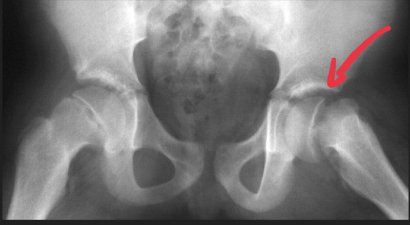
Slipped Capital Femoral Epiphysis, is a common condition of the proximal femoral physis that leads to slippage of the metaphysis relative to the epiphysis, and is most commonly seen in adolescent obese males.
Diagnosis can be confirmed with radiographs of the hip.
Treatment is usually percutaneous pin fixation. Contralateral pinning is indicated for patients at high risk, such as those with an initial slip at age < 10, obese males, and those with endocrine disorders
A 43-year-old left-handed man with a history of type 2 diabetes mellitus and hypertension presented for evaluation of abnormal posturing and control of his left ring and little fingers. He initially denied injury; however, on further questioning, he recalled being kicked in the hand by an aggressive student while working as a school principal. This direct blow to the base of his left palm on the hypothenar eminence occurred approximately 3–4 weeks prior to onset of symptoms. He denied any sensory disturbances.
He was seen by a hand surgeon who evaluated him for possible flexor pulley rupture versus central slip incompetence. Magnetic resonance imaging of the left hand revealed no evidence of pulley rupture or flexor or extensor tendon abnormalities. Over the ensuing weeks, he began to demonstrate claw deformities of the ring and little fingers and then developed atrophy of his first dorsal interosseous and adductor pollicis muscles with weakness of grip and pinch. An electromyography and nerve conduction study was performed, and the results were consistent with injury to the distal deep motor branch of the ulnar nerve with sparing of both the abductor digiti minimi muscle and sensory involvement. He was referred to occupational therapy and had some improvement in function with the use of a lumbrical bar splint; however, his symptoms remained unchanged. He subsequently saw another hand surgeon, and no additional treatment recommendations were provided.
The patient presented to our hand surgery clinic 6 months after injury. Previously obtained radiographs of the left hand were reviewed, which revealed heterotopic bone arising from the volar base of the little finger metacarpal extending proximally toward the hook of hamate . Computed tomography scan of the left hand was then obtained, which confirmed the presence of abnormal bone formation near the deep motor branch of the ulnar nerve. This raised concern for posttraumatic compressive neuropathy of the deep motor branch of the ulnar nerve caused by heterotopic ossification.
Community chat: https://t.me/hamster_kombat_chat_2
Website: https://hamster.network
Twitter: x.com/hamster_kombat
YouTube: https://www.youtube.com/@HamsterKombat_Official
Bot: https://t.me/hamster_kombat_bot
Last updated 11 months, 1 week ago
Your easy, fun crypto trading app for buying and trading any crypto on the market.
📱 App: @Blum
🤖 Trading Bot: @BlumCryptoTradingBot
🆘 Help: @BlumSupport
💬 Chat: @BlumCrypto_Chat
Last updated 1 year, 4 months ago
Turn your endless taps into a financial tool.
Join @tapswap_bot
Collaboration - @taping_Guru
Last updated 11 months, 3 weeks ago